Echocardiogram
An echocardiogram (sometimes also called a Transthoracic echocardiogram) is a non-invasive ultrasound scan that produces still or moving images of all four chambers of the heart. High frequency sound waves are sent through a device on the patient’s chest to project an image onto a monitor for a real time health assessment. The test helps to assess problems with the heart valves, chambers of the heart and the hearts ability to pump blood.
You should allow 45 minutes for this test to be undertaken.
Before your procedure:
- Take your usual medication, unless your doctor or hospital has advised otherwise and bring a list of your medications to the hospital.
- Read and follow any other instructions or information given to you by your doctor/hospital.
- Wear a two piece outfit on the day so you can easily undress to your waist.
- Please do not apply lotions, perfumes, powders or neck jewellery to ensure good skin contact of the transducer.
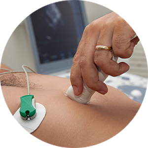
The Sonographer will discuss the echocardiogram with you prior to commencing the ultrasound. They will place ECG dots on your chest; these are also used to monitor your heart rate and rhythm during the test.
The Sonographer will put some cool gel on your chest and then move a transducer across your chest to gain pictures of your heart. They may at times ask you to briefly hold your breath.
After the procedure you may see your cardiologist for a consult to discuss the outcome of the echocardiogram – this will depend on your referrers request. A formal report will also be sent to your referring doctor and/or GP.